I hope you're doing well and enjoying the beginning of winter. Below is an article about
asbestos identification and quantification by
Alana Graham. I hope you find this article interesting and helpful.
With best wishes,
Dave Gallup
Asbestos Identification and Quantification in Bulk Samples
By Alana Graham, EMLab P&K Asbestos Analyst
What is asbestos?
Asbestos is a fibrous mineral silicate with a crystalline structure that comes from
a mineral rock. It is a great building material because it has high tensile strength,
is very flexible, and is resistant to chemicals, high temperatures and stress. As a
result, it can be found in buildings in many places including floor tiles, mastics,
caulking, roofing material, joint compound, plaster, pipe insulation, popcorn ceiling,
stucco, some wall textures, and vermiculite insulation. It is estimated that thirty
million tons of asbestos building materials were used in the U.S. between 1900 and 1975
with probably eighty to ninety percent of that still in place. This is why it can be
found in many commercial and residential buildings built before 1975. Little did we
know then that this amazing addition to building material would cause serious long
term health problems. Because of its needle-like shape, an asbestos fiber can stick
to lung tissue if inhaled and cause an inflammation. This can lead to many internal
health problems such as asbestosis, mesothelioma, lung cancer or interstitial fibrosis.
There are six types of asbestos fibers. Chrysotile, the most widely used asbestos fiber,
is the only one in the serpentine group of phyllosilicates (sheet silicates). It has a
crystalline structure that is very different from the rest. The remaining five, amosite,
crocidolite, anthophyllite, tremolite and actinolite, are all amphiboles.
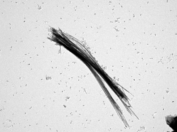
Fig. 1. Chrysotile asbestos fiber from Swift Creek viewed in a Transmission Electron Microscope (TEM).
Source: U.S. Environmental Protection Agency
How does a microscopist identify asbestos in bulk material?
The presence of asbestos in bulk building material can be detected by three methods of
microscopy. The most popular method is by Polarized Light Microscopy (PLM). With this
method, a small amount of the sample is crushed and placed on a microscope slide using
the appropriate refractive index oil. Polarized light is used to observe the many optical
properties of asbestos fibers that allow the microscopist to distinguish it from
non-asbestos fibers. A polarizing filter on a light microscope is used to force light to
vibrate in one particular plane. The polarizing filter, along with other additions to
the light microscope, such as the analyzer, a 530nm waveplate, and a dispersion staining
lens, allow the microscopist to observe properties of light and crystals such as
pleochroism (the phenomenon of substances showing different absorption colors under
transmitted light when viewed in different vibration directions), the sign of elongation
(describing the relation between the principal vibration directions and the length of
the substance), the extinction angle (the angle at which no light passes through the
substance under crossed polars), the refractive indices of the fibers (the ratio of the
velocity of light in a vacuum to the velocity of light in a substance) and birefringence
(the numerical difference between the two refractive indices of a substance). Nonetheless,
the first property observed is the morphology of the fiber. Asbestos analysis by PLM is
the least expensive and quickest method to identify and quantify asbestos fibers in
friable and non-friable material. However, there are two other methods that can produce
a more accurate quantification.
Scanning Electron Microscopy (SEM) and Transmission Electron Microscopy (TEM) are also
used to identify and quantify asbestos in a bulk sample. Both microscopes use a beam of
electrons from a filament in a vacuum. The SEM produces an image of the topography of the
sample. The electron beam interacts with the atoms on the surface of the sample and
information on the sample's composition can be collected. The TEM's electron beam passes
through the sample and an image is displaced onto a screen. Using an energy dispersive
X-ray (EDX) and a computer system, information about the fiber's elemental properties
can be gathered and graphed in their appropriate ratios. Knowing the exact ratios of the
elements in the fiber, along with other analytical information, allows the microscopist
to distinguish one type of asbestos fiber from another and asbestos fibers from
non-asbestos fibers. Selected Area Electron Diffraction (SAED) is another component of
the TEM that allows for a differentiation of fibers. It allows the microscopist to
observe the diffraction patterns of the crystalline structures of the fiber being analyzed.
The advantage of using an SEM for asbestos analysis is it has better resolution than
the PLM. As a result, the sample can be observed at higher magnifications and at a
greater depth of focus. TEM, which is more widely used than SEM, has the best
resolution out of the three microscopes and therefore is mandated for analysis of
asbestos in water. Using the TEM, asbestos sample composition can be observed at
magnifications of up to 19,000x, as opposed to the PLM which allows for a maximum
magnification of 400x, or the SEM which can be used at magnifications of up to 5,000x
for asbestos analysis. The higher magnification used by the TEM allows for more
accurate asbestos concentration estimations in samples that contain very little
asbestos (less than 5%). In addition, more accurate identifications and estimations
are attributed to the gravimetric reduction of non-friable organically bound bulk
samples selected for TEM analysis. That is, TEM samples are ashed at 480°C for
6-8 hours to eliminate the organic binder and/or tar material, and are then treated
with acid for a few minutes to eliminate the carbonate material. This allows hidden
asbestos fibers to surface and be easily noticed and identified under the TEM.
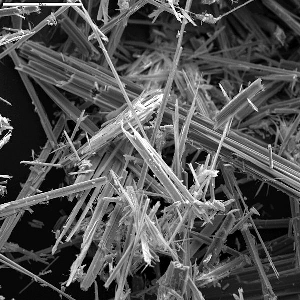
Fig. 2. Anthophyllite asbestos, Georgia.
Source: U.S. Geological Survey Denver Microbeam Laboratory
How do microscopists quantify asbestos in a bulk sample?
Calibrated visual estimation (CVES) is the quantification method widely used in
asbestos analysis via PLM, SEM, and TEM. Initially, as they are being trained to
quantify asbestos in a sample, microscopists use reference slides/samples of known
concentrations and compare with reference documents having photomicrographs to
learn how to estimate asbestos percentages. The microscopists continue to estimate
percentages in different samples until the lab manager/trainer deems them proficient
in quantification. Then, the microscopists view reference slides daily, analyze
proficiency samples, analyze monthly unknown samples, and perform inter- and
intra-laboratory analyses and other comparisons to test the continual accuracy of
their CVES. Having well trained analysts employing good quality control processes
is the best way to identify and quantify asbestos consistently and accurately.
Another method of quantification is the point count method. This method is only used
with PLM, and usually at a magnification of 100x. In this method, a special reticule
is used in the microscope allowing the microscopist to count fibrous materials that
land under a point of reference in the reticule. Multiple preparations of the sample
are made on a slide, covered with a cover slip, and then point counted. A point count
allows for lower detection limits than CVES offers. A
200 point count
has a detection limit of 0.5%, a
400 point count
of 0.25% and a
1000 point count
of 0.1%. This method is seemingly more accurate, yet it is not highly recommended
because in some cases, asbestos-containing materials do not have the same asbestos
concentrations in every area of the sample and thus different preparations will yield
results that are inconsistent and imprecise. Nevertheless, when this method is requested,
the microscopist is expected to thoroughly mix and homogenize the sample so as to evenly
disperse the asbestos so the point count estimation will be as accurate as possible.
In TEM analysis, the CVES of asbestos concentration in the gravimetrically reduced sample
is multiplied by the percentage of sample remaining after ashing and acid treatment to give
the estimation of asbestos in the entire sample. The same method applies to a PLM
gravimetric point count
where the samples are gravimetrically reduced and then point counted using PLM. The point
counted percentage is multiplied by the percentage of sample remaining after ashing and
acid treatment to give the estimation of asbestos in the entire sample.
References:
1. Oklahoma Department of Labor: Asbestos Background and History
2. McCrone, Walter C. 1987. Asbestos Identification. Chicago: McCrone Research Institute.
3. Purdue University: Scanning Electron Microscope
4. De Stefano, Luca. 2002. SEM Quantitative Determination of Asbestos in Bulk Materials.
Microscopy and Analysis 16(3): 13-15.